Back Muscles Anatomy / Back Muscles Anatomy Chart 744 1140 Anatomy System Human Body Anatomy Diagram And Chart Images. The muscles of the lower back help stabilize, rotate, flex, and extend the spinal column, which is a bony tower of 24 vertebrae that gives the body structure and houses the spinal cord.the spinal. The surface muscles of the upper back include the trapezius muscles (traps) and posterior deltoids. They provide movements of the spine , stability to the trunk, as well as the coordination between the movements of the limbs and trunk. These muscles determine body posture and also regulate the three basic movements of the trunk: The human spine is composed of 4 sections of vertebrae.
The muscles of the back can be arranged into 3 categories based on their location: This blog post article is an overview of the muscles of the lumbar spine of the trunk. These muscles include the large paired muscles in the lower back, called erector spinae, which help hold up the spine, and gluteal muscles. Both the deltoid and the trapezius are firmly attached to the spine of the scapula. The deltoid, teres major, teres minor, infraspinatus, supraspinatus (not shown) and subscapularis muscles (not shown) all extend from the scapula to the humerus and act on the shoulder joint.
3d Rendered Medically Accurate Illustration Of The Deep Back Muscles Canstock from comps.canstockphoto.com The back consists of the spine, spinal cord, muscles, ligaments, and nerves. Both the deltoid and the trapezius are firmly attached to the spine of the scapula. The multifidus, a long muscle that travels nearly the entire length of the back.it helps to stabilize and rotate the lower back, and additionally takes some. The muscles of the lower back help stabilize, rotate, flex, and extend the spinal column, which is a bony tower of 24 vertebrae that gives the body structure and houses the spinal cord.the spinal. Anatomy of the back muscles the latissimus dorsi muscles (also known as the lats) are the largest muscles of the back. The superior part of the appendicular skeleton that includes clavicle, scapula, and humerus, is attached to the axial skeleton that consists of skull. Together these muscles form a column, known as the erector spinae. What are the lower back muscles and their anatomy?
These muscles give height and breadth to back development.
The extrinsic back muscles are located in the back, but act to produce movements of the shoulder and assist respiration. Male body major muscles, flat cartoon vector style infographic illustration. We think this is the most useful anatomy picture that you need. Back pain is common and might be caused by a problem with a muscle. Muscle or ligament strains can occur from repeated use of the muscles, or from improperly or awkwardly lifting heavy objects. They start at the top of the neck and go down to the tailbone. These muscles give height and breadth to back development. Leaning back to straight vertical and all points in between. Anatomy of back muscles your back consists of three distinct layers of muscles, namely the superficial layer, the intermediate layer, and the deep layer. The muscles of the back muscles make up a large part of the anatomy (structure) of the back. Three types of back muscles that help the spine function are extensors, flexors and obliques. The muscles of the back can be arranged into 3 categories based on their location: Fit athlete lifting weight with blue muscle light concept on background.
The surface muscles of the upper back include the trapezius muscles (traps) and posterior deltoids. The deep muscles develop in the back called intrinsic muscles. Your lower back (lumbar spine) is the anatomic region between your lowest rib and the upper part of the buttock. Together these muscles form a column, known as the erector spinae. Balance the weight of your head on top of your spine evenly distribute weights from your upper body into the lower extremities
Superficial Back Muscles 1831 Artwork Stock Image C014 7820 Science Photo Library from media.sciencephoto.com We hope this picture anatomy of back muscles diagram can help you study and research. The muscles of the lower back help stabilize, rotate, flex, and extend the spinal column, which is a bony tower of 24 vertebrae that gives the body structure and houses the spinal cord.the spinal. The extrinsic back muscles are located in the back, but act to produce movements of the shoulder and assist respiration. These structures work together to support the body, enable a range of movements, and send messages from the brain to the. Back muscles the muscles of the back are a group of strong, paired muscles that lie on the posterior aspect of the trunk. Balance the weight of your head on top of your spine evenly distribute weights from your upper body into the lower extremities They are short muscles associated with the spinous and transverse processes of the vertebrae. 1 your spine in this region has a natural inward curve.
These layers of back muscles help to mobilize and stabilize your trunk during your day to day activities.
The deep muscles develop in the back called intrinsic muscles. Superficial muscles of the back are located directly deep towards the skin along with superficial fascia.they are occasionally called the appendicular group as these muscles are mainly associated with activities of the appendicular skeleton. These layers of back muscles help to mobilize and stabilize your trunk during your day to day activities. These sections are cervical (neck), thoracic (upper and middle back), lumbar (lower back), and sacrum (tailbone). Superficial back muscles, intermediate back muscles and intrinsic back muscles.the intrinsic muscles are named as such because their embryological development begins in the back, oppose to the superficial and intermediate back muscles which develop elsewhere and are therefore classed as extrinsic muscles. For more anatomy content please follow us and visit our website: The back muscles are divided into two large groups: (2017, elsevier) should be consulted. These muscles include the large paired muscles in the lower back, called erector spinae, which help hold up the spine, and gluteal muscles. The extensor muscles are attached to back of the spine and enable standing and lifting objects. Muscles of the back anatomy. These muscles give height and breadth to back development. The human spine is composed of 4 sections of vertebrae.
The muscles, bones, ligaments, and tendons in the back can all be injured and cause back pain. Anatomy chart courtesy of fcit the latissimus dorsi muscles (also known as the lats) are the largest muscles of the back. They provide movements of the spine , stability to the trunk, as well as the coordination between the movements of the limbs and trunk. The human spine is composed of 4 sections of vertebrae. The deep muscles develop in the back called intrinsic muscles.
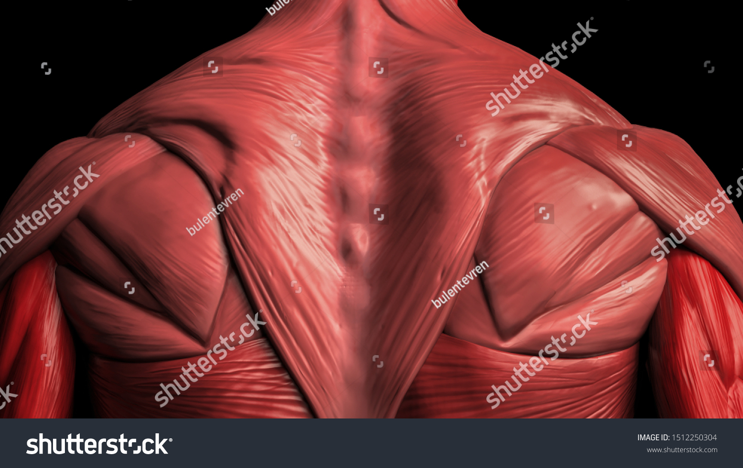
Back Muscles Anatomy Male 3d Render Stock Illustration 1512250304 from image.shutterstock.com The back muscles are anatomically layered into superficial (extrinsic) and deep (intrinsic) muscles. The abdominal muscles also play a major role in the posture and stability to the body and compress the organs of the abdominal cavity during various activities such as breathing and defecation. The anatomy of the back muscles basically defines their functions according to the location they occupy in the human body and to the other structures with which they connect. (2017, elsevier) should be consulted. They provide movements of the spine , stability to the trunk, as well as the coordination between the movements of the limbs and trunk. Anatomy chart courtesy of fcit the latissimus dorsi muscles (also known as the lats) are the largest muscles of the back. We hope this picture anatomy of back muscles diagram can help you study and research. The muscles of the back can be arranged into 3 categories based on their location:
The three deep muscles of the back include the semispinalis, multifidus, and rotatores.
The multifidus, a long muscle that travels nearly the entire length of the back.it helps to stabilize and rotate the lower back, and additionally takes some. The deep back muscles are posterior to the erector spinae. Three types of back muscles that help the spine function are extensors, flexors and obliques. The trapezius and latissimus dorsi muscles connect the upper limb to the vertebral column. Anatomy of back muscles your back consists of three distinct layers of muscles, namely the superficial layer, the intermediate layer, and the deep layer. The extrinsic back muscles are located in the back, but act to produce movements of the shoulder and assist respiration. The anatomy of the back muscles basically defines their functions according to the location they occupy in the human body and to the other structures with which they connect. Leaning back to straight vertical and all points in between. We think this is the most useful anatomy picture that you need. The intrinsic back muscles are found deeper to the extrinsic muscles, separated from them by the thoracolumbar fascia. Muscle or ligament strains can occur from repeated use of the muscles, or from improperly or awkwardly lifting heavy objects. To perform clinical clinical orthopedic manual therapy to the lumbar spine. Deep muscles of the lower back include:
Share :
Post a Comment
for "Back Muscles Anatomy / Back Muscles Anatomy Chart 744 1140 Anatomy System Human Body Anatomy Diagram And Chart Images"


Post a Comment for "Back Muscles Anatomy / Back Muscles Anatomy Chart 744 1140 Anatomy System Human Body Anatomy Diagram And Chart Images"